EV71 Brainstem encephalomyelitis: Pathological findings in Malaysia
From our experience with 4 autopsies of patients who died of Enterovirus 71 infection in the University Hospital, Kuala Lumpur, Malaysia, (see Cases 1 & 2, and Cases 3 & 4) we found that the pathological changes were mainly confined to the brainstem and spinal cord.
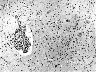
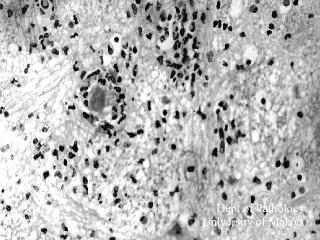
The typical histopathological findings in the brainstem and spinal cord did not differ much from other types of viral encephalitides, and consisted of perivascular inflammation, microglial nodule formation, neuronal necrosis and phagocytosis, and mild meningitis. No neuronal inclusions were detected. The extent of the inflammation was remarkable in that it involved all levels of the spinal cord, medulla, tegmentum of the pons (spares the basis pontis) and much of the midbrain (spares the peduncles). In our cases there was no apparent involvement of the cerebellum, cerebrum or myocardium.
Figure 1 : Medulla: Perivascular inflammation and inflammatory cells in the parenchyma.
Figure 2: Medulla: Intense inflammation with oedema, microglial nodule formation, and phagocytosis. Some neutrophils are also noted.
- Note: Click on image to see a larger picture
- Source:
- Dr K T Wong (WONGKT@medicine.med.um.edu.my)
- Assoc. Professor,
- Dept of Pathology,
- University of Malaya,
- 50603 Kuala Lumpur
- Malaysia
- 18 June 1998


