In December 2021, Philip O'Keefe, an Australian who was living with amyotrophic lateral sclerosis (ALS), posted a message on Twitter through the account of Synchron's chief executive. The remarkable part was not the message itself, but how he sent it. As ALS progressively deprived him of the use of his hands, an implanted device enabled him to select computer commands using signals recorded from his brain.[1]
The device, called the Stentrode, had been placed through a blood vessel rather than through an opening in his skull. For a patient living with severe paralysis, its value lay in something straightforward: a new way to communicate and exercise some independence.
How does it work? What do clinical studies tell us about safety? And, most importantly, does it improve patients' lives?
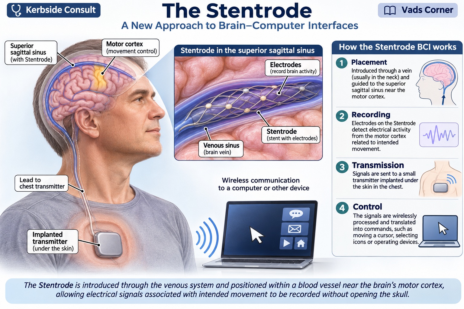 AI-generated illustration
Contents
- What is a brain–computer interface?
- From laboratory experiments to clinical applications
- The Australian Stentrode
- What have the clinical studies shown?
- Safety and side effects
- Quality of life — beyond the technology
- How does it compare with other innovations?
- From research to routine clinical care
- What lies ahead?
- At the kerbside
- Sources & further reading
What is a brain–computer interface?
When we intend to move, networks in the brain generate electrical activity. A brain–computer interface (BCI) detects certain patterns of that activity and translates them into commands for a computer or another device. A patient who cannot physically move a hand may still produce a recognisable signal when attempting the movement.
This is not unrestricted mind-reading. The system is trained to recognise particular patterns associated with particular tasks. It may also work alongside eye tracking or other assistive technology; not every cursor movement or letter selection is controlled by the implant alone.
From laboratory experiments to clinical applications
Key milestones in brain–computer interfaces
| Period | Milestone |
|---|---|
| 1920s | Hans Berger records human electroencephalographic activity (first recording 1924, published 1929), establishing a foundation for non-invasive brain recording.[2] |
| 1973 | Jacques Vidal describes the brain–computer interface concept.[3] |
| 1988 | Farwell and Donchin demonstrate a P300-based letter-selection approach in healthy volunteers.[4] |
| 1998 | Kennedy and Bakay report a locked-in patient with ALS who learns to control signals recorded from an implanted electrode in her brain, an early step towards computer control.[5] |
| 2006 | The BrainGate pilot trial reports a man with tetraplegia using signals from a 96-electrode array in his motor cortex as a "neural cursor" to open simulated email and operate devices such as a television.[6] |
| 2012 | Researchers demonstrate brain-controlled robotic-arm reach-and-grasp tasks in two people with tetraplegia.[7] |
| 2016 | Australian investigators publish foundational endovascular Stentrode research in sheep, with recordings for up to 190 days.[8] |
| 2019–2023 | The SWITCH study (started 2019) implants four patients and reports 12-month safety and feasibility findings in 2023.[9] |
| 2024–2026 | COMMAND reports interim safety findings in a conference abstract.[10] Other implanted interfaces also enter human testing (see “How does it compare with other innovations?” below). |
The emphasis has moved from proving that brain signals can control a machine to establishing whether patients can use these systems reliably at home for years.
The Australian Stentrode
Developed through research involving the University of Melbourne and Synchron, the Stentrode comprises an expandable nitinol mesh carrying 16 platinum electrodes. It is delivered by catheter through the jugular vein and positioned in the superior sagittal sinus near the motor cortex. Signals pass through an implanted lead to a chest unit, which communicates with external equipment.[8], [9], [11]
The approach avoids a craniotomy and penetration of brain tissue. It does not avoid invasive treatment altogether: vascular access, an implanted lead and a chest unit are still required. Nor should it be described as a simple outpatient procedure for every patient. In SWITCH, implantation took place under general anaesthesia and averaged about 232 minutes, including testing of the device during the operation; three of the four patients went home within 48 hours and one stayed a further day.[9]
What have the clinical studies shown?
The peer-reviewed Australian SWITCH study analysed four implanted patients with severe paralysis. A fifth patient had been enrolled but was withdrawn before implantation because pre-implant MRI showed isolated drainage through one transverse sinus. All four implanted patients completed 12 months of follow-up and demonstrated computer control. The primary safety endpoint was device-related serious adverse events resulting in death or permanent increased disability; the investigators reported no serious adverse events, no vessel occlusion and no device migration.[9]
All four were men of European ancestry, chosen for preserved motor cortex activity and suitable venous anatomy. The authors themselves note that the absence of serious events in an ALS cohort reflects careful patient selection: adequate lung function, respiratory support where needed and no comorbidities at enrolment. The results may therefore not transfer to every patient. All four initially used eye tracking to move the cursor, with the implant providing the click; one later used the implant alone to type, answer yes/no questions and send text messages.[9]
The American COMMAND study enrolled six participants. Its registered primary outcome was treatment-related serious adverse events at 12 months.[12] In a 2024 conference abstract, investigators reported no serious adverse events resulting in death or permanent increased disability, and no sinus occlusion, stenosis or device migration, at the time of abstract preparation. Two participants had not yet completed 12-month follow-up. The abstract also lists quality-of-life measures among its pre-specified secondary outcomes, but does not report them.[10] Synchron later announced that all six participants met the primary endpoint, but that is a company statement reported in the trade press, not a peer-reviewed result.[13] For now, COMMAND is best read as interim, partly company-reported evidence.
A subsequent analysis of the ten participants from both studies (eight with ALS, one with primary lateral sclerosis and one with a brainstem stroke) found considerable variation in motor signal strength between individuals. The strongest predictor was how far the device overlapped the primary motor cortex, which underlines the importance of precise placement.[11] This is a preprint, not yet peer reviewed, and its authors caution that it should not be used to guide clinical practice.
One further point deserves mention. SWITCH was funded by Synchron, which also took part in the study's design, conduct, analysis and manuscript, and several authors disclosed founder, shareholder or patent interests in the company.[9] The preprint's authors include Synchron employees who hold stock options, and it reports that COMMAND was supported by the US National Institutes of Health and SWITCH by Synchron.[11] None of this invalidates the findings, but readers should know who generated them, and independent replication will matter.
In short, the evidence supports feasibility and short-term safety in a handful of carefully selected patients. It does not yet show lasting benefit or comparative superiority.
Safety and side effects
The most relevant potential risks are bleeding, injury to the vein, venous thrombosis, infection, implanted-lead or transmitter failure and complications of antiplatelet treatment. Tissue incorporation around a vascular implant may aid fixation, but creates questions about removal or replacement.
In SWITCH, the investigators reported eight mild adverse device effects, without a serious event meeting the primary endpoint during 12 months. These included headache and bruising at the incision sites, and they resolved without intervention. The authors also describe one patient with a haematoma at the jugular puncture site, without mass effect and without need for surgical drainage.[9] This is encouraging, but four patients cannot establish the frequency of uncommon complications. Two of the four participants later died of complications of ALS, one month and 12 months after completing the study, which the investigators judged unrelated to the device.[9] Even the pooled early experience of about ten implanted participants is too small to establish long-term safety, or any safety advantage over intracortical implants.
Patients received antiplatelet treatment as part of the clinical protocol: dual antiplatelet therapy from two weeks before implantation, continued for three months, followed by aspirin for at least a year.[9] The duration and suitability of such treatment must be assessed individually; it should not be assumed that an implant can simply be inserted and forgotten.
Selected references: 9.
Quality of life — beyond the technology
For someone with profound paralysis, being able to express discomfort, make a choice, contact a friend or communicate privately with family may be more important than a laboratory measure of decoding accuracy. These are matters of autonomy, dignity and participation.
Yet we should not assume that severe physical disability necessarily means poor quality of life, or that an implant automatically improves it. Patients' own goals and perceptions must guide assessment. Meaningful outcomes include communication accuracy and speed, fatigue, psychological well-being, social participation, satisfaction, caregiver workload and the ability to use the system independently at home.
These outcomes need to be measured prospectively with suitable patient-reported instruments, not inferred from impressive demonstrations. The burden of training, maintenance, clinical visits and technical support must also be counted. A useful system should improve daily life by more than the burden it imposes.
One long-term participant illustrates the point. Rodney Gorham, an Australian with ALS, has lived with his Stentrode since December 2020. A March 2026 WIRED report described his progression from simple selections to controlling a wider range of computer and household functions, alongside increasing fatigue and concentration demands as ALS progressed. The account drew on interviews with his wife, a Synchron clinical engineer and the company’s CEO; it should be read as reported experience rather than a validated quality-of-life outcome.[14]
His experience shows that successful implantation is not the same as sustained benefit: as ALS progresses, a patient's ability to operate a device can change even when the device itself works.
The evidence so far is descriptive. In SWITCH, satisfaction was reported informally by patients under the care of the referring investigator, and the benefits were patient-specific: faster communication for one patient who previously used eye tracking alone, contact with friends and family overseas for another, and the ability to organise a music collection for a third.[9] Synchron's own website describes the system as investigational and states that its clinical benefit has not been validated.[15]
How does it compare with other innovations?
How the main implanted-interface programmes compare
| Programme | Approach | Principal aim |
|---|---|---|
| Synchron Stentrode | Electrodes in a cerebral vein | Digital communication and control |
| Neuralink N1 | Threads inserted into brain tissue[16] | Digital and external-device control |
| Paradromics Connexus | Intracortical microelectrode arrays[17] | Speech and communication |
| Precision Layer 7 | Thin-film electrodes on the brain surface[18] | High-density recording |
| Neuracle NEO | Epidural electrodes above the dura[19] | Assisted hand movement |
| BrainGate | Intracortical research arrays[6] | Communication and external-device control |
Neuralink, associated with Elon Musk, has attracted public attention, but it is only one branch of a much longer research history. Intracortical systems may capture more detailed signals, while endovascular systems avoid penetrating brain tissue. More electrodes do not necessarily mean a better outcome for the patient.
Regulatory status also differs. Precision’s array received US FDA 510(k) clearance in April 2025 for implantation of up to 30 days,[18] China approved Neuracle’s NEO system for commercial use in March 2026,[19] and Paradromics reported its first permanent Connexus implant in its Connect-One study in June 2026.[17] These programmes also have different intended uses, so a single numerical ranking would be misleading. A system designed to restore speech should not be judged by the same performance measures as one intended to control a robotic glove.
From research to routine clinical care
Several questions remain: who is suitable for implantation, how long does the benefit persist, who pays for ongoing technical support, and what happens if the device fails or its manufacturer stops supporting it? Patients who become dependent on an implanted communication system should not be left without a continuity-of-care plan.
Non-invasive options, particularly eye tracking and conventional assistive communication devices, remain essential comparators. For some patients they may provide adequate benefit without surgical risk.
The Stentrode remains investigational,[15] and to my knowledge it is not currently available in Malaysia. The immediate priority remains access to multidisciplinary ALS care, rehabilitation and assistive communication. Any future adoption of implanted BCIs would require regulatory oversight, specialist capacity, patient selection, funding and long-term support.
Selected references: 15.
What lies ahead?
The Stentrode is only one approach in a rapidly developing field. Researchers are exploring ways to make brain–computer interfaces more capable, easier to use and less dependent on specialised laboratory support. The objective is not simply to demonstrate that a computer can respond to brain signals, but to give people with severe disabilities greater independence in everyday life.
 AI-generated illustration
Restoring speech and facial expressions
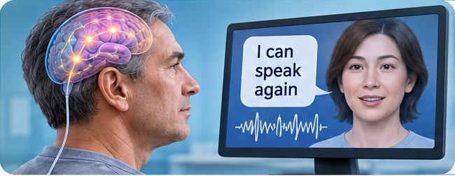
Experimental systems are being developed to translate brain activity associated with attempted speech into words. Others aim to animate digital avatars with facial expressions. For someone who has lost the ability to speak, these approaches may eventually allow more natural communication than selecting individual letters on a screen. The landmark 2023 demonstration involved a single participant with a brainstem stroke and a 253-electrode array on the surface of the brain.[20] A 2025 report from the same group, in a participant with severe paralysis and anarthria, used surface recordings to drive a streaming speech synthesiser personalised to her pre-injury voice, decoding in 80-millisecond increments, which reduces the delay that disrupts natural conversation.[21]
Illustration of an experimental speech-decoding interface; not a photograph of a clinical result.
 AI-generated illustration
Reconnecting the brain with paralysed muscles
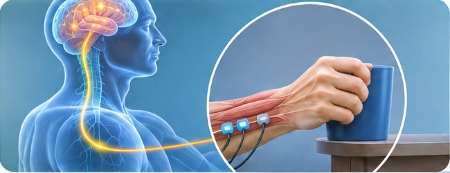
Other researchers are investigating ways to link brain signals to electrical stimulation of muscles or the spinal cord, with the aim of restoring selected movements. In a 2017 report, one man with a high cervical spinal cord injury used signals from two intracortical arrays to command stimulation of his own arm and hand muscles.[22] In 2023, a brain–spine interface linked cortical signals to stimulation of the spinal cord in one participant with tetraplegia, who could stand and walk in community settings.[23] A 2026 report described a "double neural bypass" combining an intracortical interface with spinal and cortical stimulation in one man with chronic complete tetraplegia; it reported self-feeding, manipulation of delicate objects, and persistent gains in elbow flexion and wrist tactile sensation.[24] Early demonstrations are encouraging, but each involved a single participant and they cannot yet be considered established treatments.
Conceptual illustration of brain-guided stimulation for hand movement.
 AI-generated illustration
Brain-controlled walking and rehabilitation
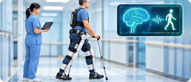
Brain-guided exoskeletons, as in a single-patient proof-of-concept report in 2019, and brain–spine interfaces are being explored as ways to help people practise movement.[25], [23] In the 2023 brain–spine study, the participant could walk overground with crutches even when the interface was switched off, suggesting a rehabilitation effect.[23] A 2026 study went further by adding artificial leg sensation: in one epilepsy patient with temporary implanted electrodes, not a person with spinal cord injury, decoded stepping intent drove a robotic exoskeleton while stimulation of the sensory cortex produced leg percepts. It establishes feasibility only.[26] These approaches remain experimental: assisted movement should not be confused with a cure for paralysis or a return to independent walking.
Illustration of an exoskeleton used in a supervised rehabilitation setting.
Less dramatic, but equally important, challenges include reliable implants over many years, simple home use, affordability and continuing technical support. Whether these advances lead to sustained improvements in independence, psychological well-being and quality of life will ultimately matter more than the sophistication of the technology.
For now, these developments are promising directions rather than proven solutions. They will deserve closer examination when larger clinical studies and longer follow-up become available.
At the kerbside
The Stentrode demonstrates that clinically useful brain signals can be recorded from within a blood vessel without penetrating brain tissue. Early results are promising, but larger studies and longer follow-up are needed before long-term safety, effectiveness and comparative value can be established.
Does it improve patients' lives? Individual patients describe real benefit, but formal quality-of-life results are not yet available in the evidence reviewed here.
Ultimately, success should not be measured by electrode count or software sophistication. It should be measured by whether a patient can communicate, participate in daily life and retain a degree of independence, with a benefit that outweighs the risks and burdens of treatment.